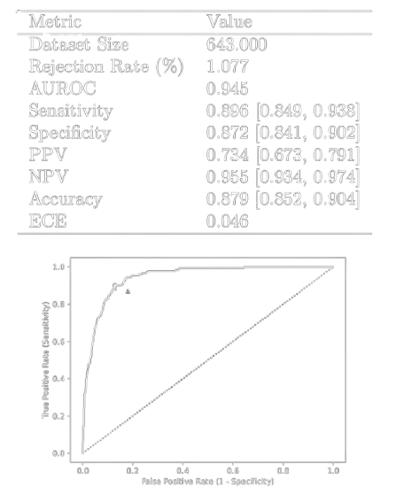
Read about updates on iCardio.ai.

Reach out to us and integrate AI into your pipeline today.

bringing ai to ultrasound

iCardio.ai solutions.

Redefining the future of heart imaging.

echomeasure™
echocheck™
cardiovision™
icardio.ai ice
autolabel ice
echomeasure™ lv
autogeo lv
echomeasure™
echocheck™
cardiovision™
icardio.ai ice
autolabel ice
echomeasure™ lv
autogeo lv
autogeo lv
echomeasure™ lv
autolabel ice
icardio.ai ice
cardiovision™
echocheck™
echomeasure™
autogeo lv
echomeasure™ lv
autolabel ice
icardio.ai ice
cardiovision™
echocheck™
echomeasure™
O
510(k) cleared solution

iCardio.ai EchoMeasure

Bringing AI precision to Echocardiography

Despite its ubiquity, echocardiography is prone to significant intra-reader variability. As a solution to this issue, AI has been extensively investigated as a means to minimize variability in quantitative interpretation of echocardiography. Artificial intelligence AI has shown superiority over human readers in the interpretation of echocardiography, including ejection fraction estimation.[4] EchoMeasure can automate over a dozen cardiac measurements and can be seamlessly integrated into the standard reporting workflow.

[4] He, Bryan, et al. "Blinded, randomized trial of sonographer versus AI cardiacfunction assessment." Nature (2023): 1-5.

Considering the ever-growing demand for echocardiograms, cardiologists and sonographers often spend valuable time adjusting and creating measurements. These repetitive tasks require the consideration of numerous parameters, and multiple button clicks. A study indicated 90% of clinical sonographers experienced symptoms of WRMSDs Work-Related Musculoskeletal Disorders).[5] To combat this, EchoMeasure can automate many of the standard linear and volumetric measurements of the heart ordinarily performed manually.

[5] Forecasting the Future of Cardiovascular Disease in the United States, AHAPolicy Statement, 2011, source: CIR.0b013e31820a55f5
EchoMeasure FDA-cleared measurements

The following measurements are currently available through EchoMeasure via the iCardio.ai API.

Parasternal Long Axis (PLAX)

Left Ventricular Diastolic Diameter (LVDd)
Left Ventricular Systolic Diameter (LVSd)
Right Ventricular Diastolic Diameter (RVDd)
Right Ventricular Systolic Diameter (RVSd)
Right Ventricular Outflow Tract Diameter (RVOTd)
Left Atrial Dimension (LAd)Posterior Wall Thickness (PWd)
Interventricular Septal Thickness (IVSd)
Left Ventricular Outflow Tract Diameter (LVOTd)
Diameter of the Aortic Annulus (AAd)
Left Atrial Dimension (LAd)
Posterior Wall Diameter of the Sinus of Valsalva (SoVd)
Diameter of the Sinotubular Junction (StJd)

Apical 4 Chamber (A4C)

Ejection Fraction (LVEF Simpson’s Biplane)
Left Ventricular Diastolic Area (LVDa)
Left Ventricular Systolic Area (LVSa)

Apical 2 Chamber (A4C)

Ejection Fraction (LVEF Simpson’s Biplane)
Left Ventricular Diastolic Area (LVDa)
Left Ventricular Systolic Area (LVSa)

Biplane (A4C + A2C)

Left Ventricular Ejection Fraction (Simpson’s method of discs)

iCardio.ai Chamber Measurements

Automated Measurement of Right Ventricular Diameter, Interventricular Septum. Left Ventricular Diameter, and Posterior Wall.

Calculate left ventricular dimension, right ventricular dimension, wall dimenstions, at systolic, diastolic and mid points in the cardiac cycle in the PLAX perspectives.

rv: 2.2cm
ivs: 1.2cm
lvdd: 5.1cm
lvsd: 3.7cm

iCardio.ai Aortic Apparatus Measurements

This model extracts features of the aortic apparatus, including the Left Ventricular Outflow Tract, the Aortic Annulus, the Sinus of Valsalva and the Sinotubular Junction.

high fidelity in sub-par and moving conditions

View Classification and Grading

This model extracts features in real-time using a mobilenet model architecture. By providing labeled features of the heart, ultrasound novices can orient themselves around the heart.

classify over 30 b-mode perspectives

Performance and Data Science

All models undergo rigorous performance testing. In this example, we can visualize performance of the model's prediction over each frame of the cardiac cycle.

Discover tailored iCardio.ai services.

Anonymization Services

Securely de-identify your DICOM data to meet regulatory compliance and facilitate research. We help you unlock the value of your data while maintaining patient privacy.

Custom Model Development

Collaborate with our AI experts to build bespoke echocardiography models tailored to your specific clinical and research objectives.

Data Annotation and Curation

Transform raw data into AI-ready assets. Our specialized team of clinical experts can accurately label, segment, and validate large datasets of medical images and clips, essential for training and refining high-performance AI models.
.face { backface-visibility: hidden; transform-style: preserve-3d; }